


CQ | 3D Segmentation of Medical Images with MONAI and UNet: A Practical Guide for Volumetric Analysis in Python
⚡ Reper CorpQuants: 3D segmentation with MONAI and UNet enables the automation and refinement of volumetric medical analysis, providing professionals with a robust and scalable solution for diagnosis and clinical research.
Artificial intelligence is transforming the way we analyze medical images, and 3D segmentation is opening new horizons for diagnosis and research. In this article, you will learn how to quickly build a complete volumetric organ segmentation pipeline on CT images, using the most advanced open-source tools in the field.
Whether you are an AI professional or a manager interested in innovation, you will find clear steps, concrete examples, and practical recommendations for implementing medical deep learning solutions in Python. Automated segmentation of anatomical structures not only optimizes clinical workflows, but also increases the accuracy and reproducibility of volumetric data analysis.

Why 3D Segmentation Matters in Medical Imaging
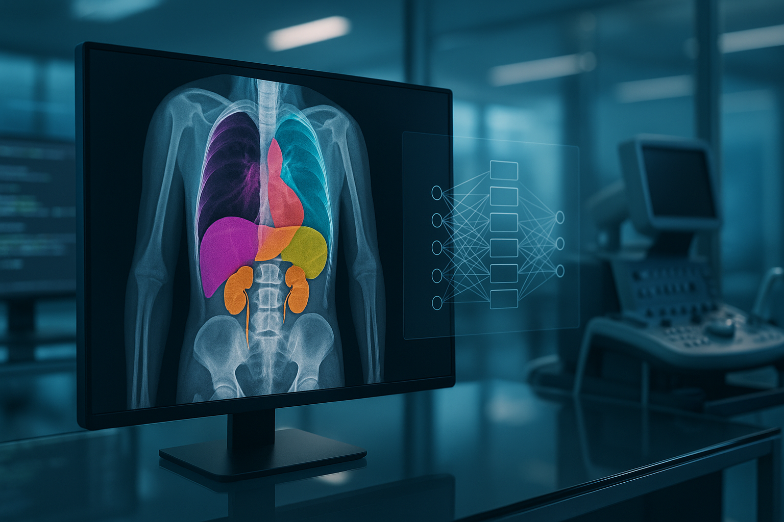
3D segmentation of medical images represents one of the most valuable applications of artificial intelligence in healthcare. By automatically identifying and delineating organs or lesions in CT or MRI volumes, 3D segmentation allows for precise evaluation of tissue structure and function, supporting diagnosis, intervention planning, and disease monitoring. Unlike 2D segmentation, 3D segmentation provides a volumetric perspective, essential for a comprehensive understanding of anatomy and pathology.
The Role of MONAI and UNet in Medical Image Processing
In recent years, the ecosystem of tools for medical deep learning has evolved rapidly. MONAI (Medical Open Network for AI) has established itself as the open-source standard for developing, training, and evaluating AI models dedicated to medical imaging. Built on PyTorch, MONAI offers specialized modules for preprocessing, augmentation, network architectures, and evaluation, optimized for volumetric data.
The UNet model remains the reference architecture for medical segmentation, thanks to its encoder-decoder design with skip connections, which enables capturing both global context and local details. In its 3D variant, UNet can process entire volumes, making it ideal for organ segmentation in CT images.
Practical 3D Segmentation Pipeline with MONAI and UNet
Implementing a complete 3D segmentation pipeline involves several essential steps, from preprocessing raw data to evaluating model performance. Here are the main steps:
1. Preprocessing Volumetric Data
- Loading and normalizing CT volumes: Use
monai.transforms.LoadImagedto load NIfTI or DICOM files, followed byScaleIntensityRangedto normalize pixel values. - Alignment and resizing: Transforms such as
SpacingdandResizedensure uniform resolution and dimensions of volumes for efficient training.
2. Data Augmentation
- Spatial augmentations: Rotations, translations, 3D flips (
RandRotate90d,RandFlipd) help increase data diversity and model generalization. - Intensity perturbations: Augmentations such as
RandGaussianNoisedorRandAdjustContrastdsimulate variations seen in clinical practice.
3. Building and Training the 3D UNet Model
- Model definition: MONAI provides
monai.networks.nets.UNetwith configurable parameters (number of channels, layers, kernel size, etc.). - Loss function:
DiceLossorDiceCELossare standard for segmentation, optimizing the overlap between prediction and ground truth mask. - Training: Use a classic PyTorch loop or
monai.engines.SupervisedTrainerto manage the forward, backward, and evaluation steps on 3D batches.
4. Evaluation and Visualization of Results
- Performance metrics: Dice Score, IoU (Intersection over Union), Sensitivity/Specificity – calculated with
monai.metrics. - Visualization: MONAI and Matplotlib allow overlaying segmentations on CT slices for rapid visual validation.
Impact and Next Steps in Adopting AI for Medical Volumetric Analysis
Adopting automated 3D segmentation with MONAI and UNet accelerates the digital transformation of medical imaging, reducing analysis time and eliminating subjective variability. Python and deep learning-based solutions can be quickly adapted to new data types and pathologies, paving the way for assisted diagnosis and large-scale clinical research.
For professionals and managers, the next steps include:
- Assessing hardware infrastructure (GPU, fast storage) for efficient volumetric processing
- Integrating AI pipelines with existing clinical workflows
- Developing customized models for specific applications (tumors, organs, rare lesions)
- Ensuring compliance with medical data security and privacy standards
(This material was assisted by an AI tool and reviewed by our team before publishing).